All specialties videos


The View of Nasopharynx after Radiotherapy Treatment...
In this video we can see the appearance of the nasopharynx some months after the radiotherapy treatment because of nasopharyngeal carcinoma. There are signs of the mucosal atrophy as a dry and thinner...

Transoral Nasopharyngoscopy Technique-The View...
Nasopharynx as a hidden part of the nose is not easily visualized. It is traditionally examined indirectly by using a postnasal mirror and reflected light. And the anterior rhinoscopic examination is...

Otoendoscopy - Appearance of The Normal Eardrum
To recognize some subtle abnormalities in certain pathologies of a eardrum it is very important to know how normal eardrum looks like. Its colour is pearly white, with a cone of light which is present...

Functional Endoscopic Sinus Surgery (FESS)- Post-Endoscopical...
In this video we can see the appearance of the left nasal cavity in a patient after the functional endoscopic sinus surgery (FESS) because of chronic sinusitis. An evidence of inferior turbinectomy, uncinectomy,...

Rima Glottis After Transoral CO2-Laser Cordectomy...
In this video we can see the appearance of rima glottidis 6 months after transmuscular CO2-laser cordectomy, which was performed because of the early stage (TI) left vocal fold cancer. There is no evidence...
The Larynx (Voicebox) - The Absence of The Epiglottis...
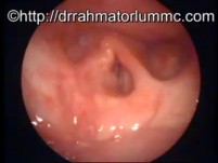
In this video we can see the endoscopic view of the larynx with an absence of its epiglottis [dotted squares]. As we can see the left vocal fold is immobile. However it is almost completely-compensated...

An Elongation of Uvula
We can associate the elongated uvula with habitual snoring or obstructive sleep apnoea syndrome (OSAS) and unexplained throat irritations and coughs. The treatment means to make it partially trimmed or...

The Vocal Fold Palsy Fully Compensated in HD
In this video we can see a fully compensated left adductor vocal fold palsy. To produce the normal voice the normal right vocal fold needs to meet the paralyzed side without phonate gap.

Middle Ear Cavity-The Hypotympanum
In this video we can see hypotympanum of the middle ear as seen through an eardrum perforation. [Black arrows - the air cells, green arrrow - the entrance into eustachian tube located anteroinferiorly]

Use of 70 Degrees Endoscope - Surface Marking...
In this video we can see the surface marking of the upper border of the thyroid cartilage as seen by using a 70 rigid endoscope. The good visualization is not possible because the overlying mucosa is...

True Vocal Folds Telangiectasia
In this video we can see telangiectasia of true vocal folds. An occurrence of haemorrhage can be either spontaneous or upon phonotrauma with the potential of scarring and dysphonia.
Obstructive Sleep Apnea Syndrome Caused by The...
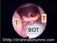
In this video we can see the changes in the airway lumen dimensions of the velopharynx and oropharyngeal areas during Müller's manoeuvre. It is routinely performed as an office evaluation of obstructive...

The View of An Adult and A Child's Eardrum
In this video we can see the normal eardrum of an adult and a child in a sequence. At birth its morphology and size are similar.

The Human Floor of Mouth and The Tongue Anatomy
In this video we can see the normal anatomy of floor of mouth and the tongue. It easy to see the ligual vein which is superficially located. It is possible to palpate bimanually the submandbular gland...

Respiratory Distress and Stridor in an Infant
There is a huge importance in recognizing signs of respiratory distress in an infant or a child. The main signs are: nasal flaring, increase breathing rate and recessions (indrawings) which may involve...


Users
-

-

Michael Hawke
Professor
-

Gottumukkala Raju
Professor
-

-

Ashley Mark
MD
Channels
-

Gastroenterology Atlas
medical institution
-

SAGES - Society of American Gastrointestinal and Endoscopic Surgeons
medical institution
-

High Impact - Medical Animation
medical institution
-

Orthopaedic Academy
medical institution
-

World Health Summit
commercial institution
Are you Health Professional?
Register now, join the community for free access.
GET ALL THE BENEFITS THAT MEDTUBE PLATFORM OFFERS:
- Unlimited access to the largest e-library of professional videos, images, publications, courses
- Connect with peers 450,000+ Healthcare Professionals from 180 countries
- Upload and share your own cases, ask questions and discuss
- Create your professional profile and build personal recognition worldwide
- Stay up-to-date with innovative techniques, treatments, guidelines, discoveries in your fields of interest; be notified
- You are informed about innovative treatments, techniques and innovations in medicine