gastroenterology videos


Duodenal B Cell Lymphoma (1 of 7)
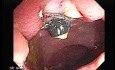
The pylorus is observed, the duodenal bulb contains several nodules.

Gastroduodenal MALT lymphoma (9 of 9)
Endoscopic view of a malignant picture involving the third part of the duodenum. H pylori infection has been associated with gastric MALT lymphomas. The role of H pylori in duodenal MALT lymphoma...

Gastroduodenal MALT lymphoma (8 of 9)
Differential Diagnosis: Gastritis or Low Grade MALT Lymphoma?. Distinguishing severe chronic gastritis from low grade MALT lymphoma is the most common diagnostic dilemma in gastric biopsies containing...

Gastroduodenal MALT lymphoma (7 of 9)
Again the video endoscopy displays several large and deep ulcers.

Gastroduodenal MALT lymphoma (6 of 9)
Endoscopy shows multiple large and deep ulcers in the third portion of the duodenum.

Gastroduodenal MALT lymphoma (5 of 9)
Endoscopy Shows large and deep ulcers The concept of mucosa-associated lymphoid tissue (MALT) lymphomas was introduced by Isaacson and Wright [ Cancer 1983; 52:1410–1416[CrossRef][Medline] in 1983....


Gastroduodenal MALT lymphoma (3 of 9)
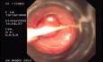
Duodenal MALT lymphoma This lesion is located in the third portion of the duodenum.

Gastroduodenal MALT lymphoma (2 of 9)
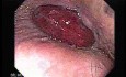
Gastroduodenal MALT lymphoma Endoscopy shows large erosion, biopsies were positive to be a maltoma. Mucosa-Associated Lymphatic Tissue (MALT) Lymphoma is a very rare type of Non-Hodgkin’s Lymphoma....

Gastroduodenal MALT lymphoma (1 of 9)
Mucosa-Associated Lymphoid Tissue Lymphoma Endoscopy shows nodular erosions with necrotic material of the gastric body and fundus as well as larger irregular ulcers of the third portion of duodenus....

Duodenal Diverticula
Pathophysiology: The cause of this condition is not known. It is believed to develop as the result of abnormalities in peristalsis, intestinal dyskinesis, and high segmental intraluminal pressures.

Duodenal Diverticula (2 of 2)
Large septated diverticulum Great majority of duodenal diverticula are asymptomatic. Clinical presentation may be characterized by non-specific abdominal symptoms and less than 5% of patients have...

Duodenal Diverticula (1 of 2)
Duodenal diverticula are acquired outpouchings of the mucosa and submucosa, 90 percent of which are on the medial aspect of the duodenum. They are rare before age 40. There is a high incidence of gallstone...

Duodenal Diverticula
Congenital diverticula are commonly found on the medial wall of the duodenum adjacent to or involving the papilla of Vater, and as a result are of particular relevance to ERCP. As the neck of the diverticulum...

Duodenal Diverticula (2 of 2)
When diverticula are located near the major duodenal papilla they are called juxtapapillary diverticula (JD) JD are important because they may obstruct the biliary and pancreatic ducts. Furthermore,...


Users
-

Gottumukkala Raju
Professor
-
-

-

-

Ahmad Hormati
Professor

Channels
-

Gastroenterology Atlas
medical institution
-

EUS - ENDO
commercial institution
-

SAGES - Society of American Gastrointestinal and Endoscopic Surgeons
medical institution
-

American Association for Primary Care Endoscopy
medical institution
-

Colonscopy Quiz by Dr. Raju
medical institution
Popular
PEG Tube Removal
Murat Akbaba
views: 37420
Prolapsed Hemorrhoid (1 of 4)
Gastroenterology Atlas
views: 33068

Double Barrel Colostomy
Mahmoud Ibrahim Ali Ali
views: 5878
Recently active users
-

EUS and more
medical institution
-

-
-
-

MKA e-class
commercial institution

Are you Health Professional?
Register now, join the community for free access.
GET ALL THE BENEFITS THAT MEDTUBE PLATFORM OFFERS:
- Unlimited access to the largest e-library of professional videos, images, publications, courses
- Connect with peers 450,000+ Healthcare Professionals from 180 countries
- Upload and share your own cases, ask questions and discuss
- Create your professional profile and build personal recognition worldwide
- Stay up-to-date with innovative techniques, treatments, guidelines, discoveries in your fields of interest; be notified
- You are informed about innovative treatments, techniques and innovations in medicine