Are you Health Professional?
Register now, join the community for free access.
GET ALL THE BENEFITS THAT MEDTUBE PLATFORM OFFERS:
- Unlimited access to the largest e-library of professional videos, images, publications, courses
- Connect with peers 450,000+ Healthcare Professionals from 180 countries
- Upload and share your own cases, ask questions and discuss
- Create your professional profile and build personal recognition worldwide
- Stay up-to-date with innovative techniques, treatments, guidelines, discoveries in your fields of interest; be notified
- You are informed about innovative treatments, techniques and innovations in medicine

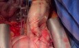
Right Lung Pneumonectomy and Arterial and Venous Stump Reconstruction in Patient with Invasive Lung Tumors

Case description
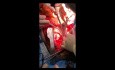
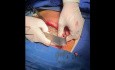
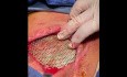
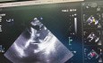
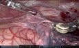
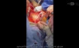
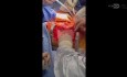
Operative Narrative: Right Pneumonectomy with Radical Vascular Reconstruction under Cardiopulmonary Bypass and Cardioplegic ArrestPreoperative Diagnosis: Malignant lung tumor of the right lung with extensive invasion into the Right Pulmonary Artery (RPA) and the right-sided superior and inferior pulmonary veins.Procedure performed: Right radical pneumonectomy, en bloc resection of invaded vascular structures, establishment of central Cardiopulmonary Bypass (CPB) via bicaval cannulation, aortic cross-clamping, anterograde aortic root cardioplegia arrest, closure of the RPA stump, and complex reconstruction of the pulmonary veins/left atrial cuff.Procedure Description1. Surgical Approach and ExposureThe patient was positioned in the left lateral decubitus position, slightly tilted posteriorly to optimize access to both the right hemithorax and the mediastinal vascular structures. A standard right posterolateral thoracotomy was performed through the fifth intercostal space. Layer by layer, the muscles were divided, and the thoracic cavity was entered. Upon initial inspection, a massive right-sided lung tumor was identified, demonstrating extensive central invasion directly into the main trunk of the right pulmonary artery (RPA) and encasing both the right superior and inferior pulmonary veins up to their junctions with the left atrium. Due to the central extent of the tumor, safe intrapericardial control of the vessels was impossible under normal beating-heart conditions. A decision was made to utilize cardiopulmonary bypass (CPB) with cardioplegic arrest to achieve radical, oncologically sound margins and safe vascular reconstruction.2. Establishment of Cardiopulmonary Bypass (Bicaval Cannulation)The pericardium was opened longitudinally anterior to the phrenic nerve and suspended with retraction sutures to expose the ascending aorta, the right atrium, and the venae cavae. Systemic heparinization was administered to achieve an activated clotting time (ACT) greater than 480 seconds.Central cannulation was performed as follows: Arterial Cannulation: A high-flow arterial cannula was placed in the distal ascending aorta. Venous Cannulation (Bicaval): To achieve total flow isolation and full access to the posterior left atrium and pulmonary vein junctions, separate bicaval cannulation was performed. The superior vena cava (SVC) was cannulated directly via a right angle cannula, and the inferior vena cava (IVC) was cannulated through the right atrial appendage.Cardiopulmonary bypass was initiated, and the patient was gradually cooled to mild hypothermia (32°C). Umbilical tapes previously looped around the SVC and IVC were snugged down tightly over the cannulae, achieving total flow isolation to completely isolate the right heart chambers and mediastinum from vsystemic venous return.3. Cross-Clamping, Cardioplegia, and ArrestAn aortic cross-clamp was applied across the distal ascending aorta. Cold blood cardioplegia was promptly delivered anterogradely through a needle root cannula into the aortic root. Prompt, complete electromechanical arrest of the heart was achieved. Topical ice slush was applied to the myocardium for additional preservation.4. Radical Tumor Resection and Vascular ReconstructionWith the heart arrested and the surgical field entirely bloodless and flaccid, the radical resection commenced: Right Pulmonary Artery (RPA) Management: The main pulmonary artery trunk was opened or visualized clearly under CPB to delineate the exact extent of tumor thrombus or wall invasion. The RPA was transected at its origin from the main pulmonary artery, ensuring a clear, tumor-free macroscopic margin. The RPA stump on the main pulmonary artery side was closed securely using a running, double-row 4-0 polypropylene suture.Pulmonary Vein and Left Atrial Cuff Resection: The tumor was found to invade the right superior and inferior pulmonary veins directly at their insertion into the left atrium. To ensure R0 resection, a generous cuff of the left atrium encompassing the orifices of both right pulmonary veins was excised en bloc with the specimen.Specimen Delivery: The remaining bronchial attachments were addressed. The right main bronchus was isolated, divided near the carina using a surgical stapler, and the entire right lung specimen along with the enclosed tumor mass and invaded vascular segments was delivered from the field. The bronchial stump was checked and found to be viable and clean.5. Left Atrial / Pulmonary Vein ReconstructionAttention was turned to reconstructing the left atrium where the large defect remained from the excised pulmonary vein cuff. Using a patch (either bovine pericardial or autologous pericardial patch, or a primary running reconstruction if tissue laxity permitted), the left atrial defect was meticulously closed. A running 4-0 or 5-0 polypropylene suture was utilized to reconstruct the smooth wall of the left atrium, ensuring no narrowing of the remaining left-sided pulmonary veins or the mitral valve inflow apparatus.6. De-airing, Weaning from CPB, and ClosureThe reconstructed left atrial cuff and aortic root were thoroughly de-aired via the aortic root vent while vent suction was maintained. The patient was placed in the Trendelenburg position. The aortic cross-clamp was removed, and sinus rhythm spontaneously restored (or restored via internal defibrillation).The patient was gradually rewarmed to 36.5°C. Once hemodynamic stability was confirmed under normal sinus rhythm, the SVC and IVC snares were released. The patient was successfully and smoothly weaned from cardiopulmonary bypass. The arterial and venous cannulae were removed, and protamine sulfate was administered to fully reverse the heparin.Excellent hemostasis was achieved across the reconstructed left atrial and pulmonary artery suture lines. Chest tubes were placed in the right hemithorax to manage post-pneumonectomy fluid balance, and the thoracotomy incision was closed anatomically in standard layers. The patient was transferred to the cardiothoracic intensive care unit in stable condition.