Are you Health Professional?
Register now, join the community for free access.
GET ALL THE BENEFITS THAT MEDTUBE PLATFORM OFFERS:
- Unlimited access to the largest e-library of professional videos, images, publications, courses
- Connect with peers 450,000+ Healthcare Professionals from 180 countries
- Upload and share your own cases, ask questions and discuss
- Create your professional profile and build personal recognition worldwide
- Stay up-to-date with innovative techniques, treatments, guidelines, discoveries in your fields of interest; be notified
- You are informed about innovative treatments, techniques and innovations in medicine

Acute Spontaneous Subdural Hemorrhage in the Subdural Space


Case description
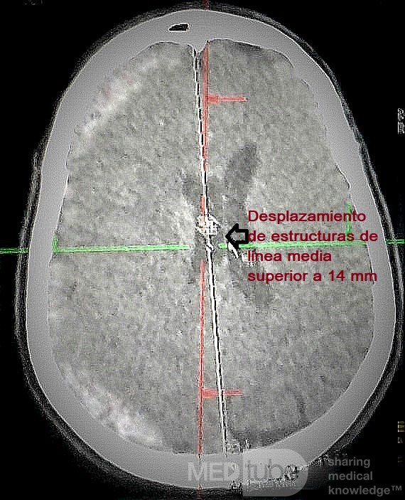
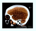
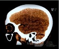
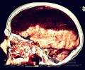
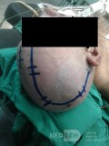
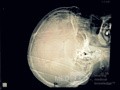
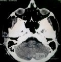
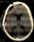
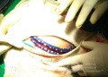
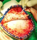
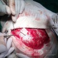
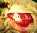
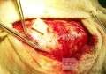
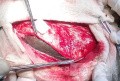
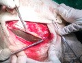
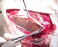
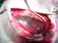
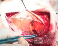
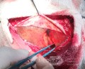
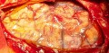
Clinical case:We present the case of a 56-year-old male patient with a history of Reactive Hepatitis who suddenly began to present headaches located in the right hemicranium for 72 hours, with difficulty walking and decreased muscle strength in the left hemibody. No history of head trauma was collected upon questioning. The multislice Computerized Axial Tomography (CT) study carried out showed a right hemispheric Acute Subdural Hematoma that displaced the midline structures by more than 5 mm with effacement of sulci and homolateral cerebral convolutions of 65 HU (Figure 1: Preoperative Multislice Skull CT Sample ). The physical examination confirmed the following signs as positive:No stigma of cranial trauma upon inspection. Acute Spontaneous Brain Hemorrhage (ASBH) or Acute Spontaneous Subdural Hematoma (ASSH) is a rare disease, generally, this type of haemorrhage is due to a traumatic history, it is produced by the accumulation of a certain volume of blood of arterial or venous origin in the space between the dura mater and the arachnoid mater in the cerebral hemisphere. Currently, the acute classification is commonly accepted for those diagnosed in the first 72 hours. An Osteoplastic Hemicraniotomy was performed, managing to evacuate the entire blood content and control the active bleeding from a cortical arterial vessel that caused it.
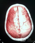
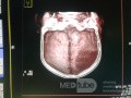
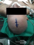
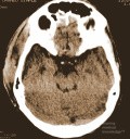
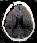
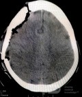
We present the case of a 56-year-old male patient with a history of Reactive Hepatitis who suddenly began to present headaches located in the right hemicranium for 72 hours, with difficulty walking and decreased muscle strength in the left hemibody. No history of head trauma was collected upon questioning. The multislice Computerized Axial Tomography (CT) study carried out showed a right hemispheric Acute Subdural Hematoma that displaced the midline structures by more than 5 mm with effacement of sulci and homolateral cerebral convolutions of 65 HU (Figure 1: Preoperative Multislice Skull CT Sample ). The physical examination confirmed the following signs as positive:No stigma of head trauma to the High blood pressure figures (160/100 mmHg).Left hemiparesis with crural predominance.Right central facial palsy grade 3 on the House-Brackman Scale.Duprés fan sign in the left lower limb, Babinski positive.Isochoric pupilsPreserved state of consciousness.Glasgow Scale for Coma 15/15 points.An extensive 10 x 12cm osteoplastic hemicraniotomy was performed, preserving the bone flap to replace it during the closure of the operation.[5-8] A Neuro-Care subdural drain coupled to a Metronic collector was placed, which was removed 48 hours later. After surgery, a simple evolutionary CT scan of the skull was performed 72 hours later, observing the presence of pneumocephalus at the right fronto-parietal level without displacement of midline structures and the absence of blood content in the subdural space. (Figure 3. Shows evolutionary CT image 72 hours after surgical treatment).The evolution of the patient was satisfactory, his status at discharge is described below:• No motor defect• No facial paralysis• Isochoric and reactive pupils• No alterations of consciousness• Glasgow Scale for Coma 15/15 points
Considerations in relation to the case:· The patient was discharged without complications.· Craniotomy in the treatment of Acute Spontaneous Subdural Hematoma allows a greater surgical field, as well as the possibility of evacuating the entire blood content, reducing ICP.· Like all surgical procedures, osteoplastic hemicraniotomy may present a risk of immediate and late intraoperative and postoperative complications that may occur (wound sepsis, intracerebral haemorrhage, subarachnoid haemorrhage, subdural empyema, edema, osteomyelitis).· All neurosurgical patients undergo a treatment process, which includes, among other pillars, surgery as the central axis and the treatment of possible complications that may appear.

Other photos of this user


Decompressive Craniotomy
Damian Lastra
views: 5001

Meningocele in an 18-year-old patient
Damian Lastra
views: 4242


Recommended


Necrosis in Echinococcosis
Nasuhi AYDIN
views: 177

Medial Clinoidal Meningioma
Nirvana Kalantari
views: 429