USG Videos - publications, events, lectures
- Street address
- Italy
- Founded at
- 2012-09-04
- Website
- USG Videos
- Views
- 21388
- Description
- This channel presents a collection of ultrasound videos.
Shared content
 26
26  0
0  0
0  0
0  0
0  0
0  0
0 
Common Femoral Vein Valve In Doppler Examination
In the following video we can observe a normal common femoral vein valve.

Severe Right Ventricle Dilatation In Echo Examination
In the following video we can observe a severe right ventricle's dilatation with unsuccessful close of tricuspidalic valve in systole. Short axis views show the septum to be flattened so that the two...

Vertebral Artery In Doppler Examination
In the following video we can observe a good quality image of vertebral artery flow.

Pericardial Effusion In Echocardiography Examination
In the following video we can observe an apical four chamber view of a two dimensional echocardiogram of a patient with pericardial effusion showing multiple fibrin strands as linear or band like structures...

Atrial Dilatation, Pericardial Effusion In Echocardiography Examination
The following material shows a case of severe bi-atrial dilatation, all valves regurgitation, pericardial effusion. Catheters in rigth chambers.

Internal Carotid Artery Occlusion In Ultrasound Exam
The following material shows a occlusion of internal carotid artery in proximal tract. On the left side there is the external carotid and on the right side there is the internal carotid artery that is...

Lower Extremity Venous Thrombosis In Ultrasound Exam
The following material demonstrates a bilateral superficial vein thrombosis of greater saphenous.


Common Femoral Vein Thrombosis And Superficial Femoral Artery Occlusion...
The following material shows a case of thrombosis of common femoral vein and occlusion of superficial femoral artery.

Mitral Valvulopathy In Echo Examination
The following material shows interestingly posterior leaflet.

Popliteal Aneurysm In Doppler Exam
The following material shows a doppler echography of the popliteal aneurysm.

Vertebral Artery, Doppler Echography
The following material shows an optimal view of vertebral artery origin (right).

Thrombosis In Right Atrium In Doppler Echocardiography
In the material we can notice a thrombosis in the right atrium in doppler echocardiography examination.

Abdominal Aortic Aneurysm In Doppler Exam
In the video we can see an abdominal aortic aneurysm, the iliac bifurcation and the aneurysm size.

Vertebral Arteries In Doppler Exam
The following material shows a small differences in size between the vertebral arteries.

Saphenous Vein Outlet In Usg Exam
The following material shows a optimal view of the saphenous vein valve.


Pleural Effusion In Echography Exam
In the video we can see a pleural effusion and bilateral lungs atelectasis.

Mitral And Aortic Stenosis In Echo Examination
The following material shows a significant calcification of mitral's posterior leaflet (all the segments with hypomobility) and of the aortic's cusps that causes a mitro - aortic stenosis.

Femoral Common Artery Bifurcation In Doppler Echography Examination
In the video we can observe the superficial femoral artery and deep femoral artery.

Atrioventricular Canal In Echocardiography Examination
In the video we can see an atrioventricular canal in Doppler echocardiography.

Ischemic Heart Disease In Echocardiography Exam
In the video we can observe the case of ischemic damage of interventricular sept and anterior wall with left ventricular dilatation and secondary mitral regurgitation. There is a pericardial and pleural...

Bilateral Pleural Effusion In Echography Examination
The following material presents a bilateral pleural effusion.

Double Left Supra Hepatic Vein View In Doppler Examination
The following material presents a suprahepatic venous system - most frequent anatomical variant.

Bicuspid Aorta View In Doppler Echocardiography
The following material presents a bicuspid aorta in ECHO.

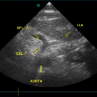
Celiac Trunk And Left Renal Vein In Doppler Examination
The following material presents an optimal view in a young man (26 years old). During the scanning there was an ectopic beat.
Nothing was found.
Are you Health Professional?
Register now, join the community for free access.
GET ALL THE BENEFITS THAT MEDTUBE PLATFORM OFFERS:
- Unlimited access to the largest e-library of professional videos, images, publications, courses
- Connect with peers 450,000+ Healthcare Professionals from 180 countries
- Upload and share your own cases, ask questions and discuss
- Create your professional profile and build personal recognition worldwide
- Stay up-to-date with innovative techniques, treatments, guidelines, discoveries in your fields of interest; be notified
- You are informed about innovative treatments, techniques and innovations in medicine
