Medial clinoidal meningiomas are among the most challenging skull base tumors, historically linked with high morbidity. Modern cranial base techniques have significantly improved outcomes. Al-Mefty Classification:
▪️ Type I – Origin below the carotid cistern → no arachnoid plane → tumor tightly adherent to the ICA & MCA → high surgical risk
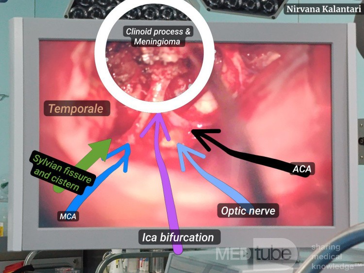
▪️ Type II – Origin above/lateral to clinoid → preserved arachnoid plane → safer dissection despite vessel encasement
▪️ Type III – Origin at optic foramen → early visual symptoms → possible lack of arachnoid plane with optic nerve
Following removal of a Type I lesion - where no arachnoid plane exists between the tumor and vessels - meticulous microsurgical technique is critical.
We use cookies to personalize content and advertising, to offer social features and to analyze traffic on our site. We share information about how you use our site with our social, advertising and analytics partners. Partners may combine this information with other data they receive from you or obtain when you use their services.
With your consent, we can better adapt our offerings to your interests and preferences.
By clicking on the "Accept all cookies" button, you declare that you consent to the installation on this device of all cookies used on our Portal by MEDtube sp. z o.o. with its registered seat at 59 Złota Street, 00-120 Warsaw ("MEDtube sp. z o.o.") and third parties, i.e. Google LLC, Meta Platform Ireland Ltd., Linkedln Ireland Unlimited Company. Due to the above-mentioned cookies, we will be able to track your behavior on the Portal and then analyze it in order to understand your use of the Portal, adapt the Portal to your needs. Cookies also allow us to remember your selected settings and personalize the Portal interface. You can find more information about the above-mentioned cookies, their functions, the entities installing them and the purposes for which they will be used in the Portal's Cookie Policy.
If you click the "Agree to Selected" button, we will use only the cookies necessary for the proper operation of the Portal, as well as those cookies that you have voluntarily agreed to collect by checking the appropriate box.
Remember that you can always specify which specific cookies will be installed on this end device. Giving your consent (by clicking "Accept all cookies") or making settings for the installation of cookies is voluntary. Once you have given your consent, you can change the rules for storing or accessing cookies at any time in the browser settings of your device. However, changing the aforementioned settings will not affect the legality of the processing of data collected through cookies installed before changing the aforementioned settings.
Cookies installed by MEDtube sp. z o.o. or information collected through them may be considered personal data under certain circumstances. The Controller of personal data is then MEDtube sp. z o.o. Contact with the Controller is possible by letter, e-mail to [email protected]. You have the right to access, rectify, delete, transfer and restrict processing of your personal data, as well as the right to lodge a complaint to the President of the Office for Personal Data Protection (ul. Stawki 2, 00-193 Warsaw). You have the right to object to the processing of your personal data. For more information on the processing of personal data on the Portal, see the Privacy Policy.
Necessary cookies ─ cookies necessary for the operation of the Portal, enabling the use of services available on the Portal.
Name
Description
Storage period
Cookie provider
PHPSESSID
Session cookie
24h
MEDtube
MT_LOGGED_IN
Stores user login information
168h
MEDtube
acceptCookies
Stores information about accepting cookie info
1 year
MEDtube
Analytical cookies ─ cookies that allow us to collect information about how you use the Service, which help us understand how our Service is used or allow us to customize it.
Advertising cookies ─ cookies that allow us to provide Service Recipients with advertising content more tailored to their preferences and interests, website visits.
Name
Description
Storage period
Cookie provider
Facebook Connect, (_fbp)
Facebook redirection information
1 quarter
Facebook
LinkedIn, (ln_or)
It is used to determine whether Oribi analyses can be performed in a specific domain